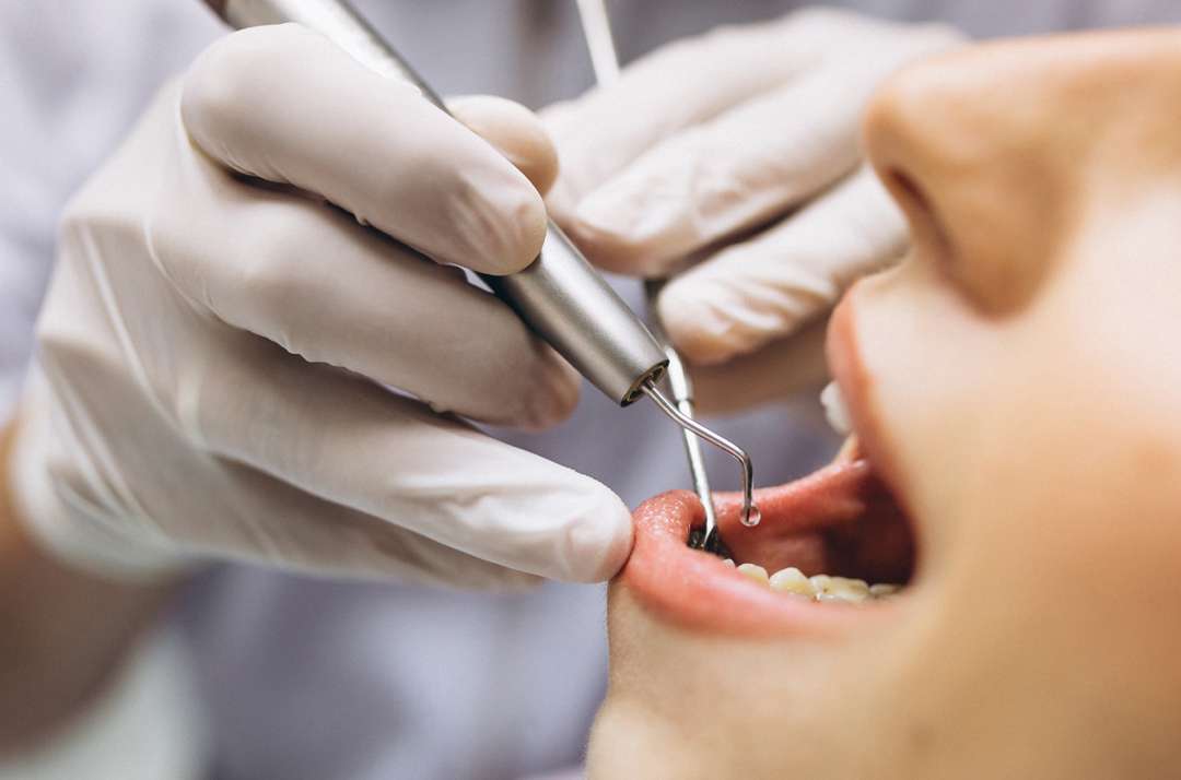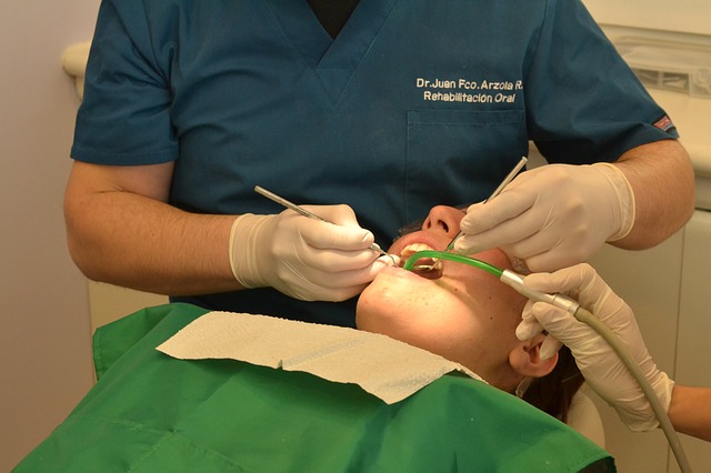
Radiography is an imaging technique using X-rays, gamma rays, or similar ionizing radiation and non-ionizing radiation to view the internal form of an object.Intraoral simply means within the mouth. There are three types of diagnostic radiographs taken in dental centers– periapical (also known as intraoral or wall-mounted), panoramic, and cephalometric. Periapical radiographs are probably the most familiar, with images of a few teeth at a time captured on small film cards inserted in the mouth.
The bisecting angle technique is accomplished by placing the receptor as close to the tooth as possible. The central ray of the x-ray beam should be directed perpendicular to an imaginary line that bisects or divides the angle formed by the long axis of the tooth and the plane of the receptor.This is commonly practiced for visualising extent of decay of tooth or to estimate bone loss occurred to teeth.
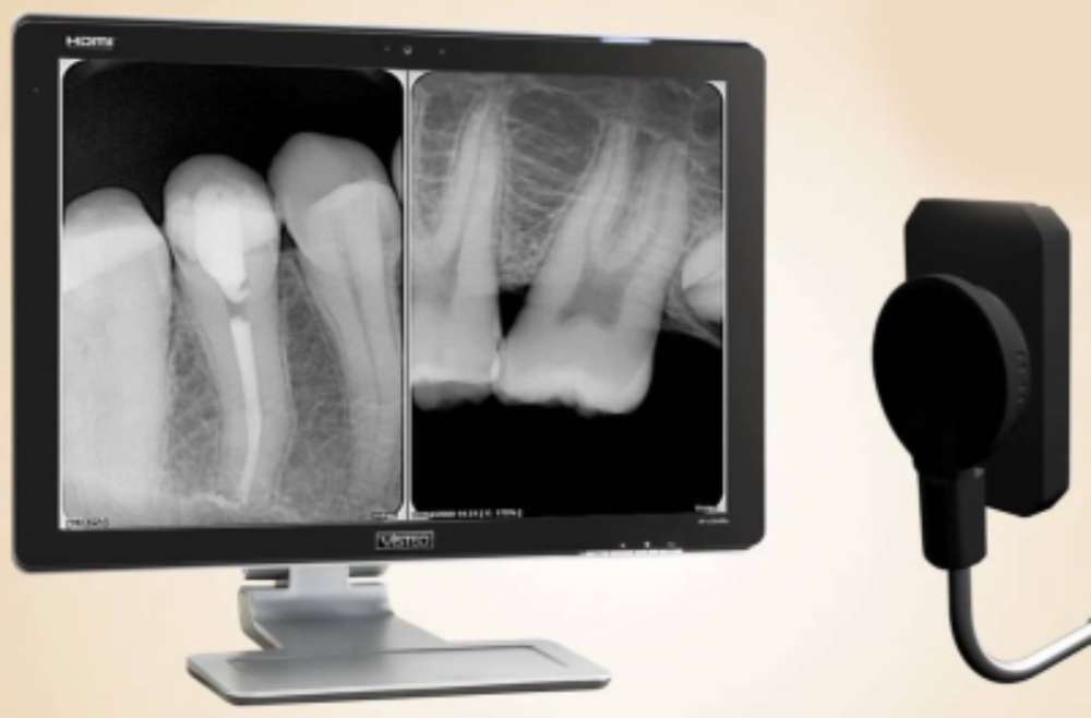
Digital radiography is a type of X-ray imaging that uses digital X-ray sensors to replace traditional photographic X-ray film, producing enhanced computer images of teeth, gums, and other oral structures and conditions. It is an advanced form of x-ray inspection which produces a digital radiographic image instantly on a computer. This technique uses x-ray sensitive plates to capture data during object examination, which is immediately transferred to a computer. This technique of radiography is practised nowadays for better visualisation of defect of teeth and to educate the patient about it and the treatment required.